Techniques
Our lab uses a range of techniques to explore changes in hippocampal structure and function. We apply methods like immunohistochemistry, electrophysiology, and biochemical assays to investigate molecular and cellular alterations. Additionally, we use genetic analysis to study how various environmental factors, such as substance exposure and brain injury, affect brain health and cognitive processes.
immunohistochemistry

Antibodies-101
Antibodies 101: How to prolong the use of antibodies in the lab.

CB1 Staining
Click here to view the CB1 Receptor immunohistochemical staining protocol.
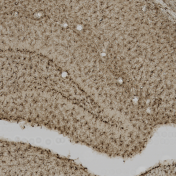
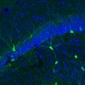
BrdU
Click here to view a video that demonstrates our immunohistochemical staining protocol for targeting BrdU positive cells in rat brain tissue. View our graphical protocol.
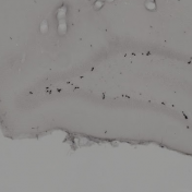
Ki-67
Graphical protocol on our immunofluorescence staining for Ki-67
Neuro-D
DCX Immunohistochemistry
DCX Immunohistochemistry on Free Floating Brain Sections
Immunohistochemistry Analysis

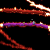
Dendritic Spine Analysis & 3D Neuron Reconstruction
Watch our video to see how we use MBF’s Neurolucida 360 software for dendritic spine analysis and 3D neuron reconstruction.
Histology

Eosin staining
Golgi-Cox Solution
Cresyl Violet
mircroscopy
Confocal Laser Scanning Microscopy
Click here to view our video series on how to use the Olympus FluoView FV1000 software.

Numerical Aperture
Watch our video on the importance of numerical aperture in microscopy.
electrophysiology
In vivo Electrophysiology

Vernier Scale
Click here to watch our video discussing how to use a vernier scale!
In vitro Electrophysiology
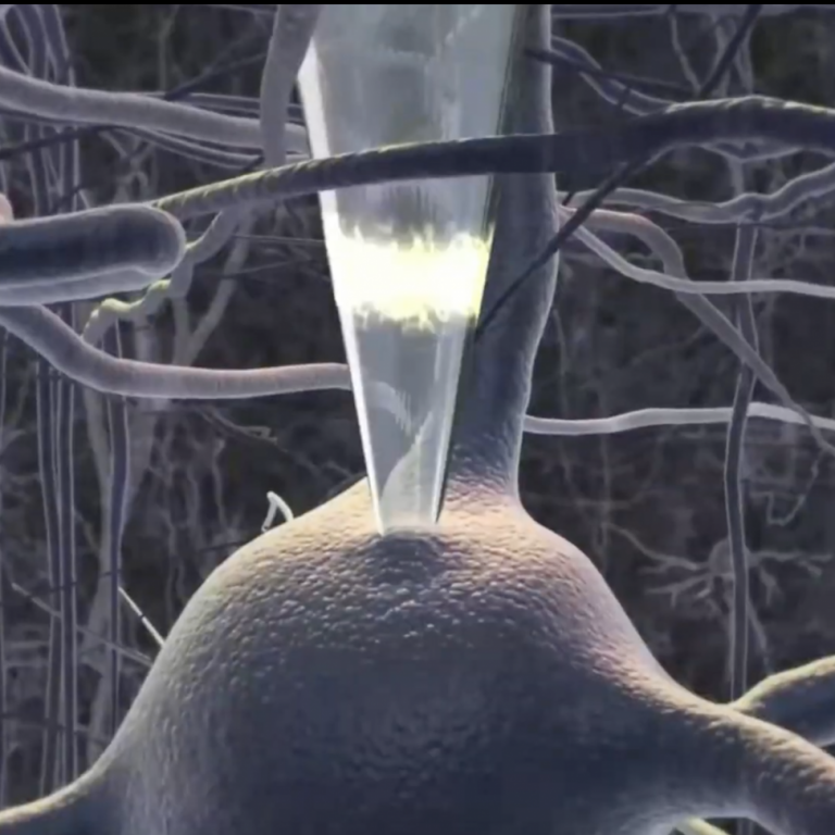
Introduction to Whole Cell
Click here to view our full video on whole cell patch clamping.

1. Preparation Steps
Steps to follow at the beginning of your experiment.

3. Slice Preparation
Click here to view our video on solution prep for whole cell patch clamping.

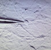
5. Finding a Cell
Click here to view our video on solution prep for whole cell patch clamping.

7. Breaking in
Click here to view our video on breaking the seal for whole cell patch clamping.
Electrode Pulling
Things to consider when pulling pipettes for recording electrodes.

2. Solution Preparation
Click here to view our video on solution prep for whole cell patch clamping.

4. Rig Setup
Click here to view our video on solution prep for whole cell patch clamping.

6. The Giga-0hm Seal
Click here to view our video on the Giga-Ohm Seal for whole cell patch clamping.
In vitro Solutions

Regular aCSF
Click here to view our video on making regular artificial cerebrospinal fluid.
Slicing & Sectioning protocols
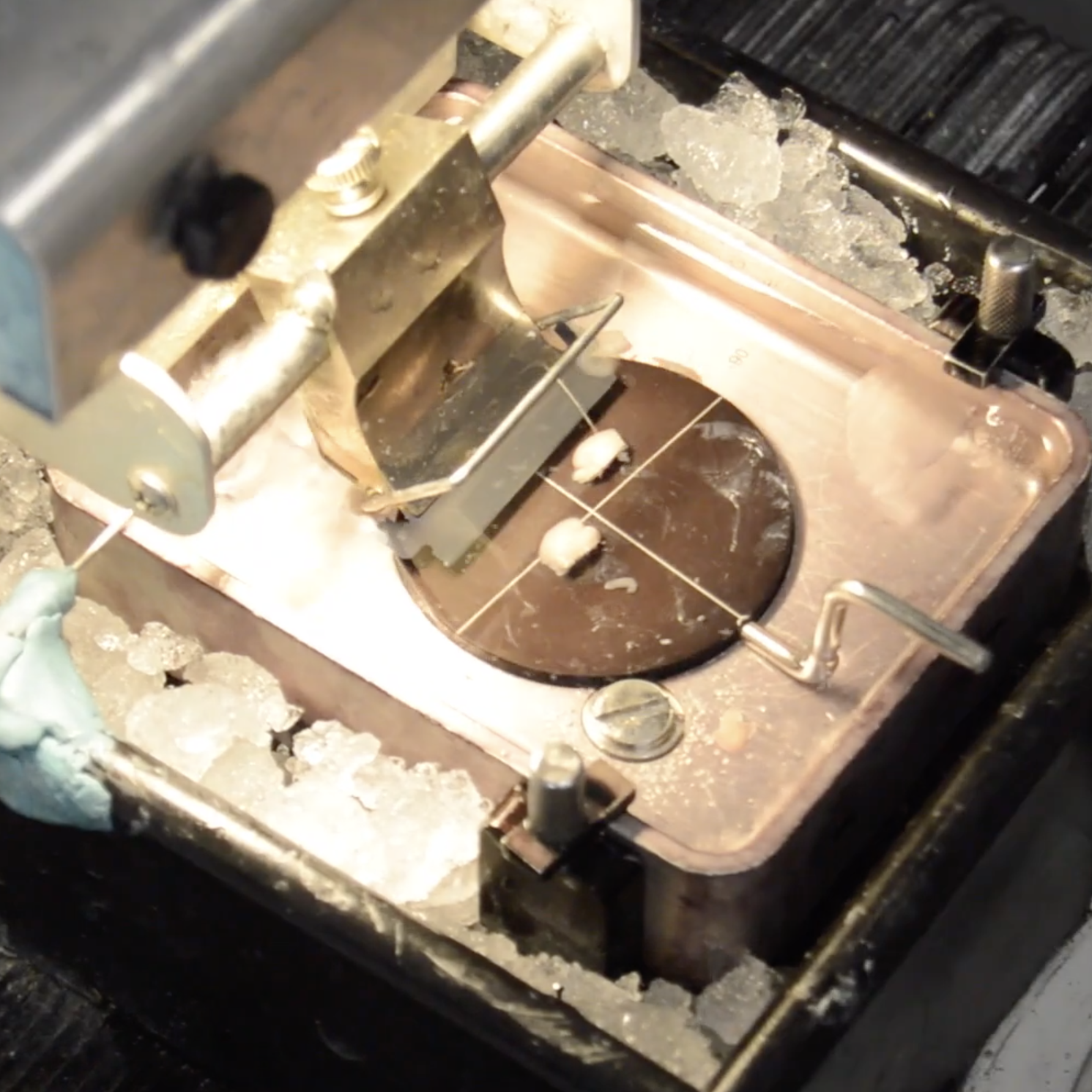
Sectioning with a Leica VT1000-S Vibratome
Click here to view our video on sectioning a mouse brain using a Leica VT1000-S Vibratome.
Sectioning with a Ted Pella 1000 Vibratome
How to section a brain using a Ted Pella 1000 Vibratome.
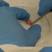
Dissection
How to dissect brain from a rat or mouse.
Sectioning with a Leica VT1000-S Vibratome
Click here to view our video on sectioning a mouse brain using a Leica VT1200 Vibratome.

Sectioning with a Precisionary Compresstome VF-310-0Z
How to section a brain into slices using a Precisionary Instruments compresstome!
Molecular
Whole Homogenate Sample Generation
View our written protocol.
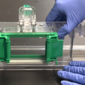
2. Gel Casting Protocol for Western Blotting
This video covers how to cast gels for western blotting. View our grapical protocol.
4. Gel Electrophoresis Protocol for Western Blotting
This video will show how to perform gel electrophoresis for western blotting. View our graphical protocol.
6. Primary Antibody Protocol for Western Blotting
This video will cover how to use primary antibodies for western blotting. View our graphical protocol.
8. Imaging Protocol for Western Blotting
This video will cover how to image a western blot. View our graphical protocol.
Synaptoneurosomes
Click here to view our flow chart.
1. Bradford Assay Protocol for Western Blotting
View our grapical protocol.
3. Sample Preparation Protocol for Western Blotting
This video will cover how to prepare samples for western blotting.
5. Transfer Protocol for Western Blotting
This video will cover how to perform the transfer protocol for western blotting. View our graphicaol protocol.
7. Secondary Antibody Protocol for Western Blotting
This video will cover how to use secondary antibodies for western blotting. View our graphical protocol.
9. Ponceau Stain Protocol for Western Blotting
This video will cover how to perform a Ponceau stain for western blotting. View our graphical protocol.
Western Blot Written Protocol
Written protocol for western blots with all steps included.
qPCR

Telomere Length Quantification
Find our video protocol and written protocol here!
Reviews, Data analysis, and statistics

PRISMA Reviews
Click here to view our video to learn how to conduct a systematic review using PRISMA.

Getting Started with R
Click here to view our video that covers the basics of R Studio, including calculating the mean, median, and standard deviation!
Prussian Blue Segmentation

Python Analysis for Electrophysiology
Our video covers a workflow for how to organize electrophysiology data for use in Python. Download Code.

Data organization and Bar Graphs in R Studio
Click here to view our video that covers how to organize cell counting data for use in R Studio, and how to make a simple bar graph!