Neuroimaging
Learn more about the imaging techniques used in our research below.
")
Magnetic Resonance Imaging (MRI)
MRI is a useful, non-invasive technique used to looks at brain structure and function in vivo. MRI can help detect these changes in healthy aging and neurodegenerative disorders. This is important for diagnosis, especially early diagnosis when brain changes precede clinical symptoms (such as in Alzheimer’s disease). It is also helpful when tracking changes to the brain over time or following treatment interventions. Since the brain is so complex, multiple MRI techniques have been developed in order to capture its systems from multiple perspectives.
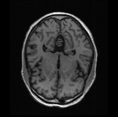
Structural MRI (sMRI)
This technique provides a snapshot of the brain with more detail than a CT scan. With contrasting brain matter, we can see abnormalities in white matter (axons) and grey matter (cell bodies). This is what most people think of when they imagine an MRI scan.
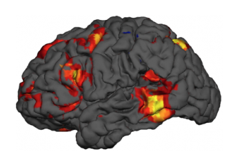
Functional MRI (fMRI)
This technique shows brain regions that are active, while performing a cognitive task or during rest. MRI detects blood-oxygen flow (aka the BOLD response) where more highly engage neural regions will require more abundant blood flow. Regions of increased BOLD can be evaluated in different healthy and patient populations to understand how brain structure relates to behaviours, or activity patterns in the default-mode network.
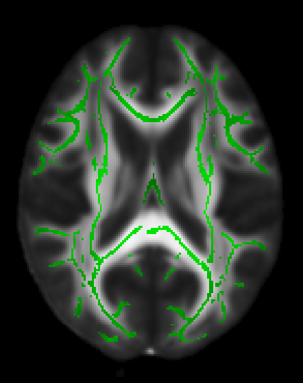
Diffusion Tensor Imaging (DTI)
This technique measures water diffusivity in the brain in order to estimate white matter integrity. This approach is used in our research to identify regions of degenerating white matter in the healthy brain and in neurodegenerative disorders, such as in MS and Alzheimer’s.
")
Functional Near Infrared Spectroscopy (fNIRS)
fNIRS is a technique that monitors brain activity using infrared light. Infrared light passes through the scull and outer most cortex and measures different regions of oxygenated and deoxygenated blood levels. Regions that show high oxygenation relative to other regions are picked up by a detector, and it can be inferred that highly oxygenated regions are particularly important for a certain task or neural mechanism.