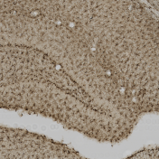
Iba1
This video covers the step by step protocol for Iba1 immunohistochemistry to target and visualize microglia.
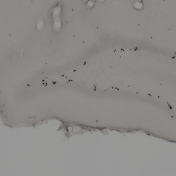
BrdU
Click here to view a video that demonstrates our immunohistochemical staining protocol for targeting BrdU positive cells in rat brain tissue. See our graphical protocol.
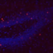
Ki-67
A video and graphical protocol on our immunofluorescence staining protocol for Ki-67 will be posted shortly – come back soon!
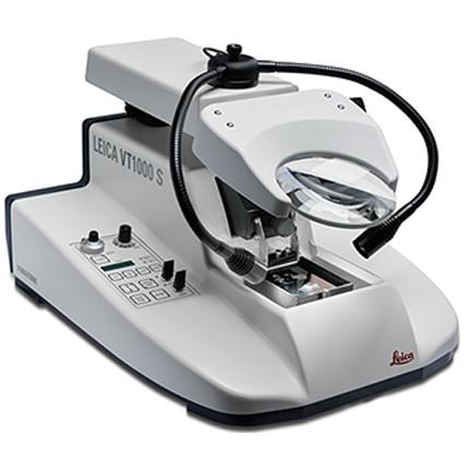
Sectioning with a Leica VT1000-S Vibratome
Click here to view our video on sectioning a mouse brain using a Leica VT1000-S Vibratome.
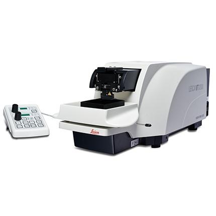
Sectioning with a Leica VT1200 Vibratome
Click here to view our video on sectioning a mouse brain using a Leica VT1200 Vibratome.
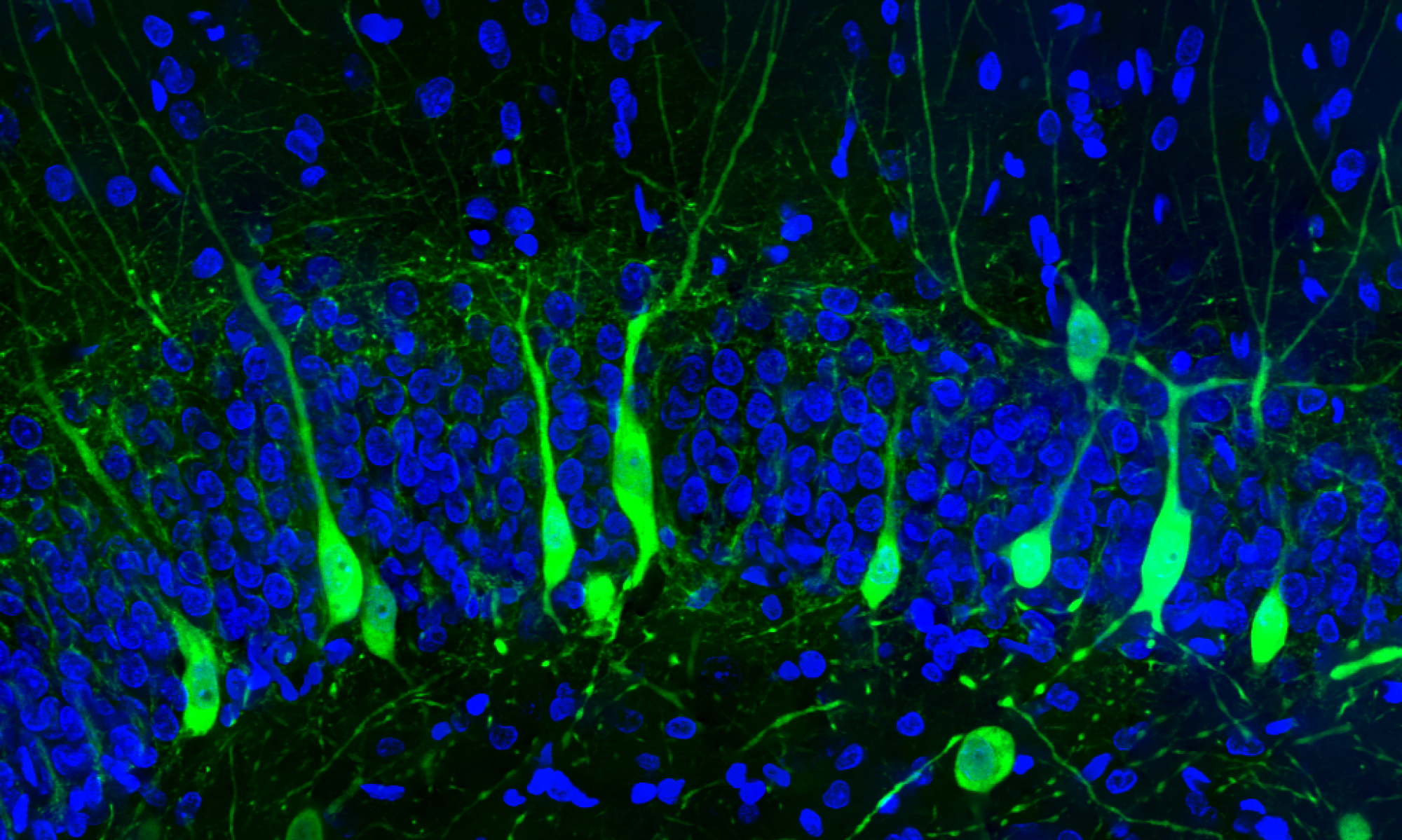
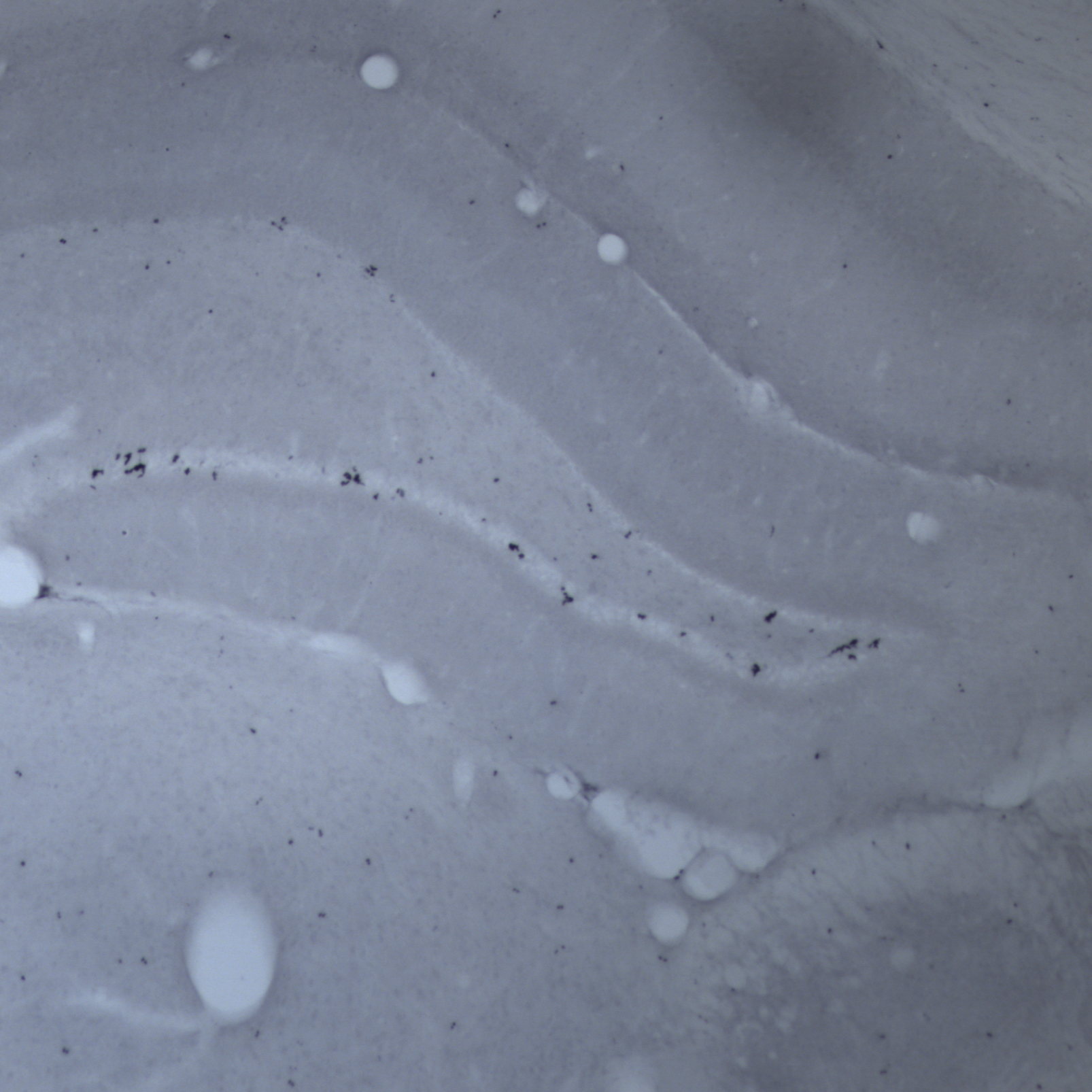
DiI For Dendritic Spine Labelling

DiI Crystal Placement and Spine Visualization
This video describes how to label neurons within the hippocampus using DiI crystals and how to visualize using confocal microscopy.
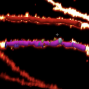
Dendritic Spine Analysis & 3D Neuron Reconstruction (Neurolucida 360)
Watch our video to see how we use MBF’s Neurolucida 360 software for dendritic spine analysis and 3D neuron reconstruction.